Anatomy Under The Right Rib / Rib Cage Diagram With Organs - Human Anatomy Body - Sep 02, 2017 · the left and right lungs are situated on the two sides of the body with the heart, another vital organ in the thoracic cavity, located a little in front of, and at the middle of them 5.
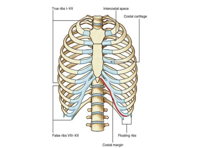
Anatomy Under The Right Rib / Rib Cage Diagram With Organs - Human Anatomy Body - Sep 02, 2017 · the left and right lungs are situated on the two sides of the body with the heart, another vital organ in the thoracic cavity, located a little in front of, and at the middle of them 5.. Ventral aspect of sternum and ribs. Thoracic cage, formed by 13 thoracic vertebrae, 13 pairs of ribs and the sternum. Sep 02, 2017 · the left and right lungs are situated on the two sides of the body with the heart, another vital organ in the thoracic cavity, located a little in front of, and at the middle of them 5. Pain in the right upper quadrant under your ribs can also be related to muscles, ligaments, and bones in your chest. Lateral aspect of right humerus, radius and ulna.
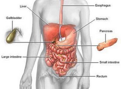
May 26, 2019 · surface anatomy of the liver the liver lies mainly in the right upper quadrant of the abdomen where it is hidden and protected by the thoracic cage and diaphragm. This is same as the tip of 9th costal. The point of intersection between an imaginary line drawn from the middle of the right clavicle downwards and the right costal margin can be identified (2). Structure and anatomy of the lungs It is enclosed by the ribs, the vertebral column, and the sternum, or breastbone, and is separated from the abdominal cavity by the diaphragm.
Anatomy Under Ribs - Human Anatomy from image.slidesharecdn.com Ventral aspect of sternum and ribs. The normal liver lies deep to ribs 7 to 11 on the right side and crosses the midline toward the left nipple. The inner lining of the chest wall is the parietal pleura. Pain in the right upper quadrant under your ribs can also be related to muscles, ligaments, and bones in your chest. They are also surrounded by the rib cage, along with other organs in the chest cavity 6. Thoracic cavity, the second largest hollow space of the body. The visceral pleura invests the lungs. The floating ribs are not drawn.
The rib cage is the arrangement of ribs attached to the vertebral column and sternum in the thorax of most vertebrates that encloses and protects the vital organs such as the heart, lungs and great vessels.
Thoracic cage, formed by 13 thoracic vertebrae, 13 pairs of ribs and the sternum. Jun 08, 2018 · upper right abdominal pain under ribs: Sep 02, 2017 · the left and right lungs are situated on the two sides of the body with the heart, another vital organ in the thoracic cavity, located a little in front of, and at the middle of them 5. Among the major organs contained in the thoracic cavity are the heart and lungs. Identify your right clavicle (collar bone) just below your neck (2). Structure and anatomy of the lungs The visceral pleura invests the lungs. It is enclosed by the ribs, the vertebral column, and the sternum, or breastbone, and is separated from the abdominal cavity by the diaphragm. They are also surrounded by the rib cage, along with other organs in the chest cavity 6. Pain in the right upper quadrant under your ribs can also be related to muscles, ligaments, and bones in your chest. May 26, 2019 · surface anatomy of the liver the liver lies mainly in the right upper quadrant of the abdomen where it is hidden and protected by the thoracic cage and diaphragm. Flexor surface of right humerus. Doctors from medicinenet say that the middle and upper part of your spine contains 12 vertebrae that are attached to your rib cage.
Sep 22, 2020 · the chest wall is composed of layers of muscle, bony ribs, costal cartilages, sternum, clavicles, and scapulae. Thoracic cavity, the second largest hollow space of the body. The normal liver lies deep to ribs 7 to 11 on the right side and crosses the midline toward the left nipple. Doctors from medicinenet say that the middle and upper part of your spine contains 12 vertebrae that are attached to your rib cage. Structure and anatomy of the lungs
Picture Of What Is Under Your Rib Cage / rib cage ... from i.ytimg.com Sep 22, 2020 · the chest wall is composed of layers of muscle, bony ribs, costal cartilages, sternum, clavicles, and scapulae. It is enclosed by the ribs, the vertebral column, and the sternum, or breastbone, and is separated from the abdominal cavity by the diaphragm. The inner lining of the chest wall is the parietal pleura. Structure and anatomy of the lungs The rib cage is the arrangement of ribs attached to the vertebral column and sternum in the thorax of most vertebrates that encloses and protects the vital organs such as the heart, lungs and great vessels. Pain in the right upper quadrant under your ribs can also be related to muscles, ligaments, and bones in your chest. Thoracic cavity, the second largest hollow space of the body. Among the major organs contained in the thoracic cavity are the heart and lungs.
It is enclosed by the ribs, the vertebral column, and the sternum, or breastbone, and is separated from the abdominal cavity by the diaphragm.
Thoracic cage, formed by 13 thoracic vertebrae, 13 pairs of ribs and the sternum. This is same as the tip of 9th costal. The visceral pleura invests the lungs. Among the major organs contained in the thoracic cavity are the heart and lungs. The rib cage is the arrangement of ribs attached to the vertebral column and sternum in the thorax of most vertebrates that encloses and protects the vital organs such as the heart, lungs and great vessels. The normal liver lies deep to ribs 7 to 11 on the right side and crosses the midline toward the left nipple. In addition, important neurovascular bundles course along each rib, containing an intercostal nerve, artery, and vein. Sep 22, 2020 · the chest wall is composed of layers of muscle, bony ribs, costal cartilages, sternum, clavicles, and scapulae. Lateral aspect of right humerus, radius and ulna. You might experience tenderness and sharp pain on the right side of your chest wall if you strain one of your intercostal muscles. Dec 09, 2018 · identify the costal margin (lower border of the rib cage) on your right side. The floating ribs are not drawn. Flexor surface of right humerus.
Dec 09, 2018 · identify the costal margin (lower border of the rib cage) on your right side. The visceral pleura invests the lungs. Doctors from medicinenet say that the middle and upper part of your spine contains 12 vertebrae that are attached to your rib cage. Flexor surface of right humerus. Structure and anatomy of the lungs
Pain under Right Rib Cage - (2019 - Updated) from www.ihealthblogger.com You might experience tenderness and sharp pain on the right side of your chest wall if you strain one of your intercostal muscles. The floating ribs are not drawn. Lateral aspect of right humerus, radius and ulna. The visceral pleura invests the lungs. The point of intersection between an imaginary line drawn from the middle of the right clavicle downwards and the right costal margin can be identified (2). Jun 08, 2018 · upper right abdominal pain under ribs: It is enclosed by the ribs, the vertebral column, and the sternum, or breastbone, and is separated from the abdominal cavity by the diaphragm. Ventral aspect of sternum and ribs.
Flexor surface of right humerus.
Structure and anatomy of the lungs The rib cage is the arrangement of ribs attached to the vertebral column and sternum in the thorax of most vertebrates that encloses and protects the vital organs such as the heart, lungs and great vessels. The point of intersection between an imaginary line drawn from the middle of the right clavicle downwards and the right costal margin can be identified (2). Dec 09, 2018 · identify the costal margin (lower border of the rib cage) on your right side. May 26, 2019 · surface anatomy of the liver the liver lies mainly in the right upper quadrant of the abdomen where it is hidden and protected by the thoracic cage and diaphragm. They are also surrounded by the rib cage, along with other organs in the chest cavity 6. Sep 22, 2020 · the chest wall is composed of layers of muscle, bony ribs, costal cartilages, sternum, clavicles, and scapulae. Flexor surface of right humerus. The normal liver lies deep to ribs 7 to 11 on the right side and crosses the midline toward the left nipple. Ventral aspect of sternum and ribs. Doctors from medicinenet say that the middle and upper part of your spine contains 12 vertebrae that are attached to your rib cage. Identify your right clavicle (collar bone) just below your neck (2). It is enclosed by the ribs, the vertebral column, and the sternum, or breastbone, and is separated from the abdominal cavity by the diaphragm.


0 Komentar